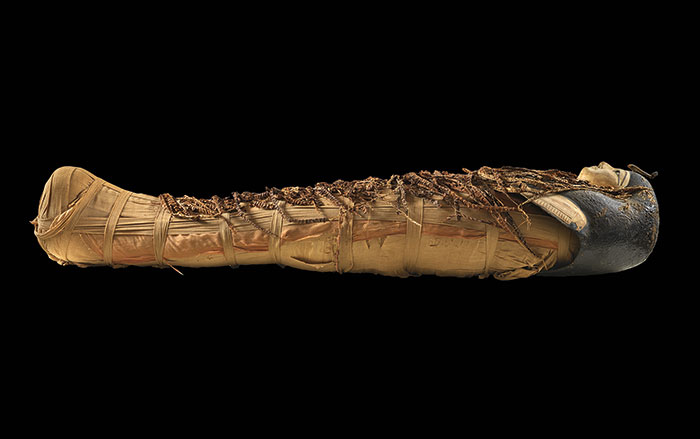
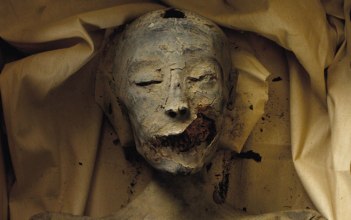
WEST PALM BEACH, FLORIDA—A team of radiologists from St. Mary’s Medical Center examined the 2,100-year-old mummy of a child from the “Tombs & Treasures of Ancient Egypt” exhibit at the South Florida Science Center and Aquarium. Based upon X-rays taken more than 40 years ago, it had been thought that the child was between the ages of four and nine at the time of death, and that she had succumbed to tuberculosis, which can wear away bone. That diagnosis relied upon what appeared to be missing vertebrae in the lower spine. (Braided hair under her gilded mask suggest the child was a girl.) Views of the girl’s teeth from the new scans indicate that she was no more than three and one-half years old at the time of death, and the missing vertebrae were found lodged in her chest. They were probably displaced during the mummification process. The doctors think that the girl died of appendicitis—a “small, bright spot” in her central abdomen is thought to be a calcified deposit that blocked the organ and caused it to rupture. “Thanks to medical science, technology, and brilliant engineering we are unlocking secrets today that can inform history more than 2,000 years old,” Lew Crampton, CEO of the South Florida Science Center and Aquarium, told The Sun-Sentinel. To read about animal mummies in ancient Egypt, see ARCHAEOLOGY's "Messengers to the Gods."
Egyptian Mummy Receives New Diagnosis
News October 17, 2014
Recommended Articles
Digs & Discoveries May/June 2024
Speaking in Golden Tongues
Rediscovering Egypt's Golden Dynasty September/October 2022
Who Was Tut’s Mother?
More to Discover
Features September/October 2014
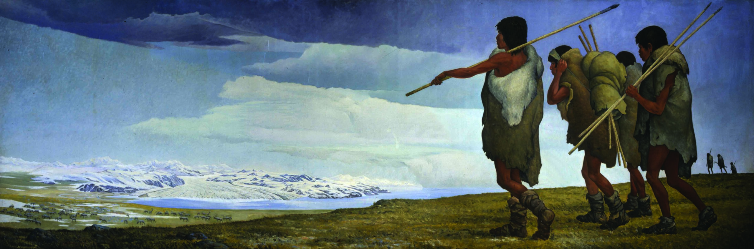
America, in the Beginning
Archaeologists continue their search for evidence of how the vast, once-uninhabited regions of the New World came to be populated
-
Features September/October 2014
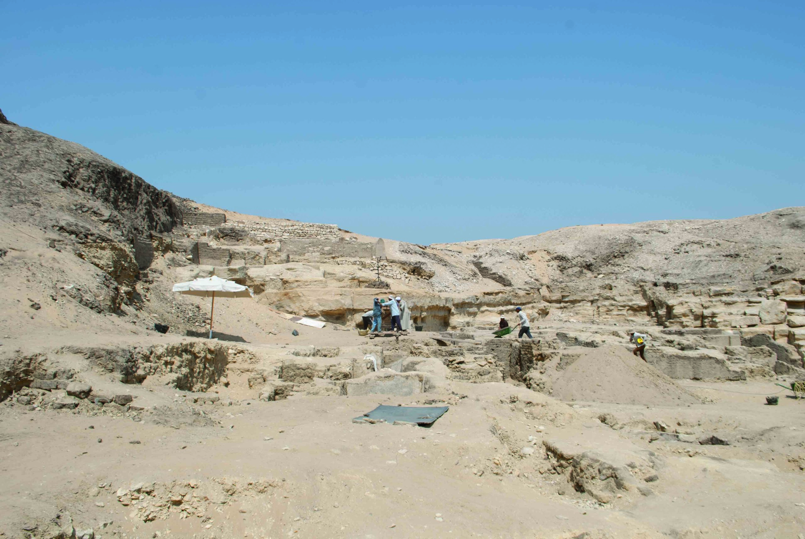
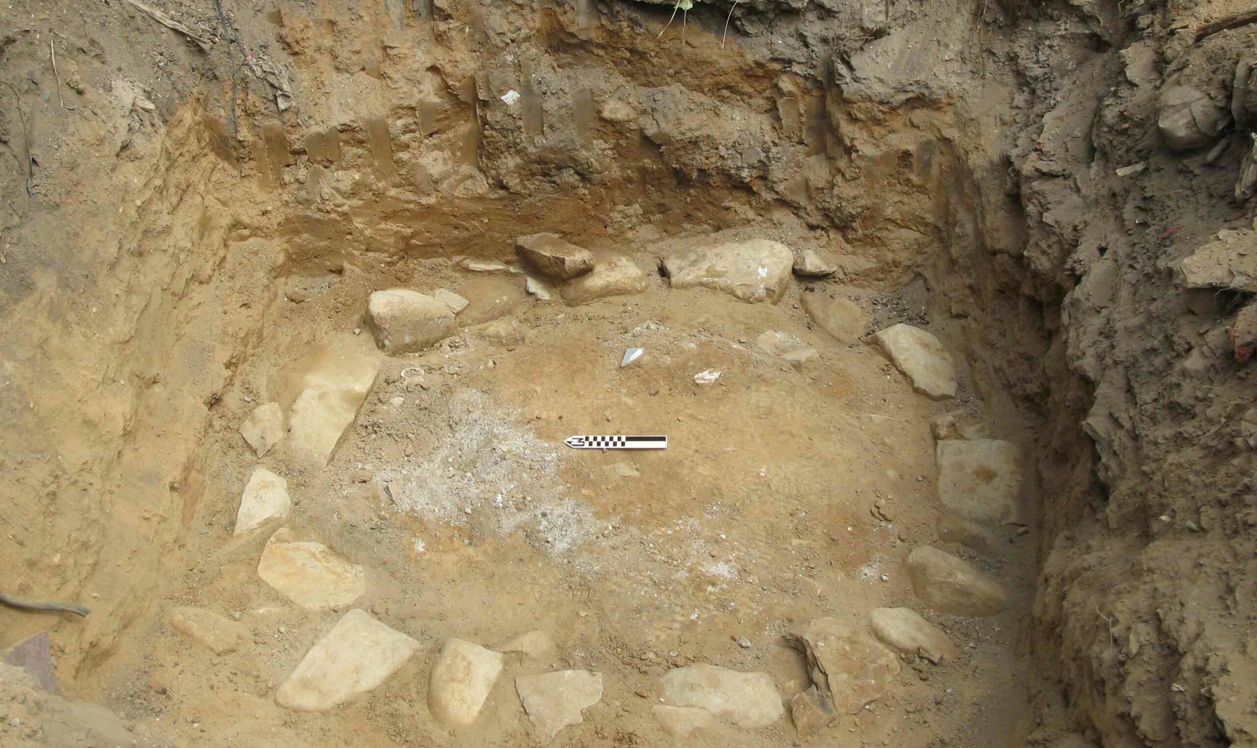
Erbil Revealed
How the first excavations in an ancient city are supporting its claim as the oldest continuously inhabited place in the world
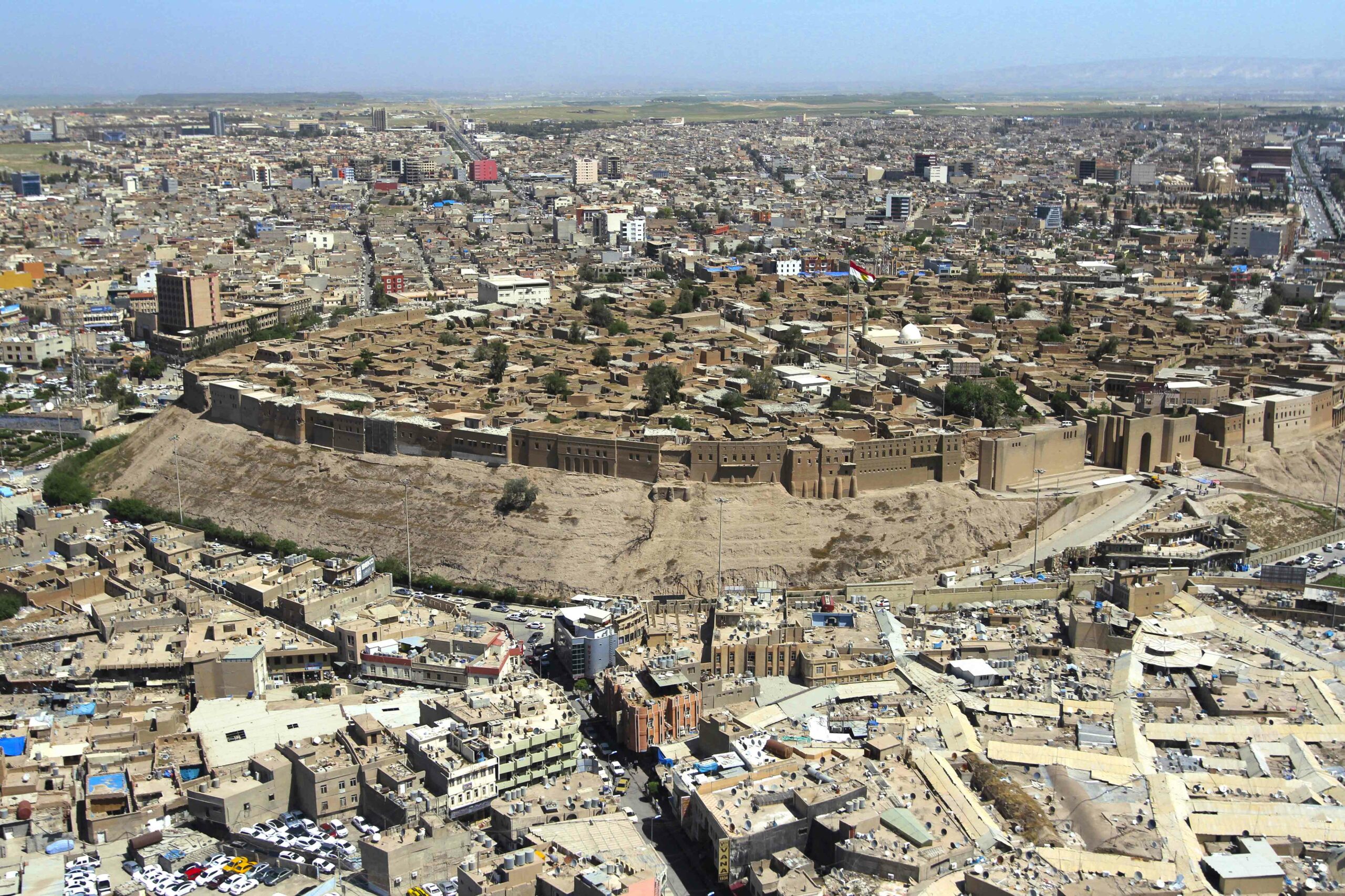 (Courtesy and Copyright Golden Eagle Global, Kurdistan, Iraq)
 -
Features September/October 2014
Castaways
Illegally enslaved and then marooned on remote Tromelin Island for fifteen years, with only ARCHAEOLOGY to tell their story
 (Richard Bouhet/ Getty Images)
 -
Letter from the Bronx September/October 2014
The Past Becomes Present
A collection of objects left behind in a New York City neighborhood connects students with the lives of people who were contemporary with their great-great-great-grandparents
 (Courtesy Celia J. Bergoffen Ph.D. R.P.A.)
 -
Artifacts September/October 2014
Silver Viking Figurine
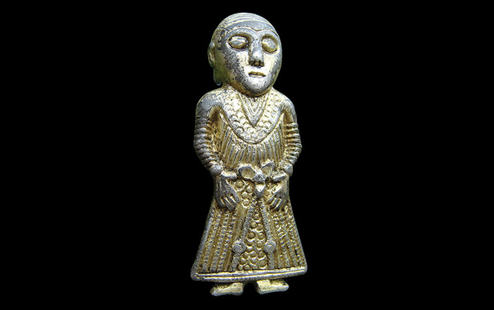 (Courtesy Claus Feveile/Østfyns Museum)