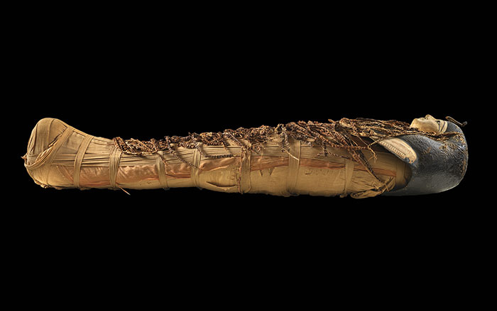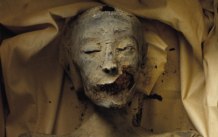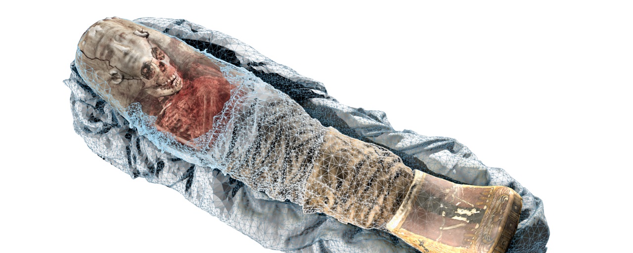
SAN JOSE, CALIFORNIA—A lifelike model of the mummy of a girl thought to have died of dysentery during Egypt’s Roman era has been created by combining computer tomography (CT) scans and 3-D scans of the mummy’s surface, according to a report in Live Science. The CT scans, taken in 2005, provide a look beneath the mummy’s wrappings, while the new surface scans, completed with a handheld 3-D scanner, captured details of its surface in color. The scans were then combined using software developed by the company Volume Graphics. The virtual model will become part of the exhibit at the Rosicrucian Egyptian Museum, where the mummy is housed. Christof Reinhart of Volume Graphics explained that the new technique could help scientists link the features on the surface of an object to associated features on the inside of an object. For more, go to “Heart Attack of the Mummies.”
New Technique Provides Thorough Look at Mummy
News November 1, 2017
SHARE:
Recommended Articles
Digs & Discoveries May/June 2024
Speaking in Golden Tongues
(Egyptian Ministry of Tourism & Antiquities)
(Courtesy Sahar Saleem)
Rediscovering Egypt's Golden Dynasty September/October 2022
Who Was Tut’s Mother?
(Ken Garrett)
(Courtesy Egyptian Ministry of Tourism & Antiquities)
-
Features September/October 2017
Painted Worlds
Searching for the meaning of self-expression in the land of the Moche
(Courtesy Lisa Trever) -
Letter from California September/October 2017
The Ancient Ecology of Fire
Lessons emerge from the ways in which North American hunter-gatherers managed the landscape around them
(Justin Sullivan / Gettyimages) -
Artifacts September/October 2017
Gilded Copper Color Disc
(Courtesy Illinois State Military Museum) -
Digs & Discoveries September/October 2017
White Horse of the Sun
(Skyscan Photolibrary / Alamy)