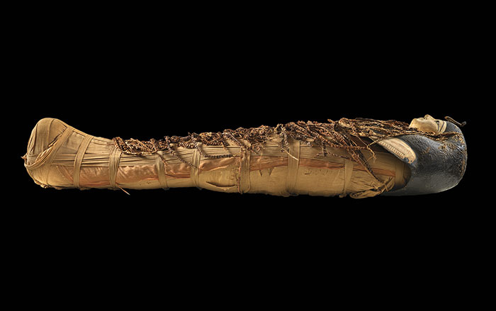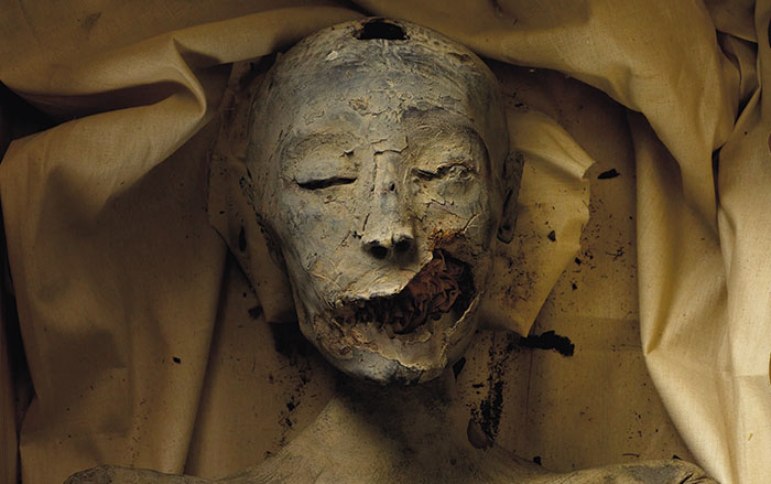

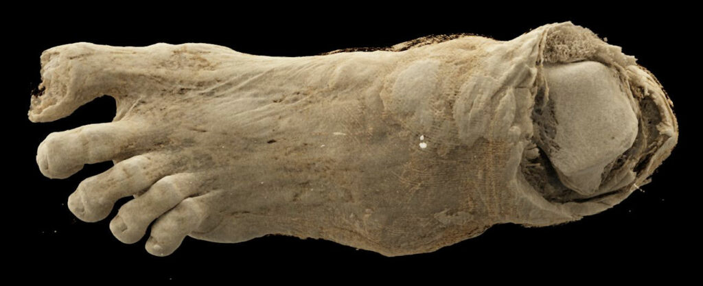
BUDAPEST, HUNGARY—Ancient Egyptian mummified remains in the collection of the MNMKK Semmelweis Museum of Medical History were examined with a CT scanner equipped with a photon-counting detector, according to a statement released by Semmelweis University. The remains in the study include two heads, two left lower limbs, a mummy bundle containing a foot, and a hand. The oldest artifacts in the collection are some 2,300 years old. The resulting images revealed the internal structure of the body parts, said team physician Ibolyka Dudás, providing a highly detailed view of abnormalities and preservation techniques used in antiquity. The new images of the lower limb suggest that the individual had osteoporosis. A second lower left limb was found to have belonged to a younger person. Images of the mummy bundle revealed layers of different types of bandages. The new information gathered could allow researchers to determine if the hand belonged to a child or an adult. “Based on the results so far, it is evident that modern imaging technology opens up new perspectives in mummy research,” said Krisztina Scheffer of the MNMKK Semmelweis Museum of Medical History. “It can reveal information hidden in finds that are thousands of years old without damaging them,” she concluded. To read about CT scanning of the sarcophagus containing the mummified body of Amenhotep I, go to "Inside a Pharaoh's Coffin," one of ARCHAEOLOGY's Top 10 Discoveries of 2022.